– OVER 12,000 SUCCESSFUL –
Tubal Reversal Procedures Performed
Since 1997


 World’s Leading Tubal Reversal Doctor
Formerly known as Chapel Hill Tubal Reversal Center, we have been performing tubal ligation reversal and corrective surgery for blocked fallopian tubes since our practice was created in 1997. We’ve pioneered innovative techniques in microsurgical tubal reversal surgery. These advancements have allowed tubal reversal to be affordable, to be performed in an outpatient setting, and not require overnight hospitalization.
We were the first doctors in the world to report within peer reviewed medical literature successful pregnancy after outpatient reversal of Essure sterilization. Since those initial case reports we have developed extensive expertise in Essure removal surgery. We offer outpatient Essure reversal and provide women who have undergone Essure sterilization an alternative to in-vitro fertilization. As a result of our extensive experience in removing Essure coils, we offer our patients having Essure symptoms a coil removal procedure with a low risk of coil fracture and provide these women with an alternative to hysterectomy.
We provide a unique surgical experience with personalized attention and one-on-one care. We make every effort to make your surgery less of a process and more of a life’s experience. Please use the buttons below for answers to our frequently asked questions.
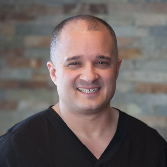
As a surgeon my reputation & integrity are extremely important to me. I’ve been providing new choices & new beginnings since 2008. I’ve had the distinct privilege of being the first surgeon in the world to report successful pregnancy within the peer reviewed medical literature after reversal of transcervical hysteroscopic tubal sterilization. Permanent is not forever at my practice! HAVE QUESTIONS?
Looking to get pregnant or repair blocked tubes? Considering a vasectomy reversal? Take the first step.
Get personalized attention and evaluations for free.

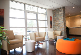
Warm and inviting our state of the art surgical facility offers safe, affordable & personalized tubal reversal surgery. Dreams of starting a family become a reality here.
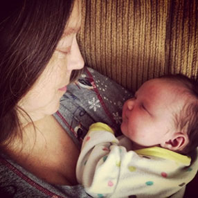
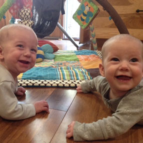 Birth Announcements
“Thank you so much for making it possible to bring these two amazing people into our lives.”
– Saratoga Springs, New York
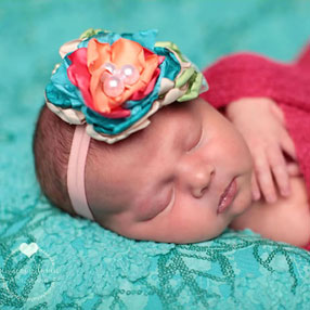
 Birth Announcements
“You are my heroes. We can never thank you enough.”
– Ontario, Canada
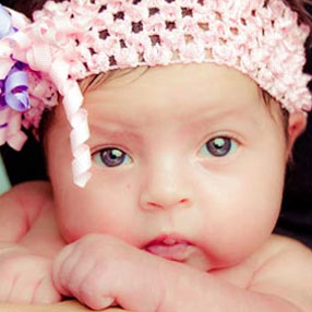
 Birth Announcements
“We have been so blessed to bring another tubal reversal baby into our family.”
– Buckatunna, Mississippi
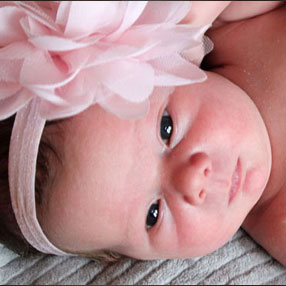
 Birth Announcements
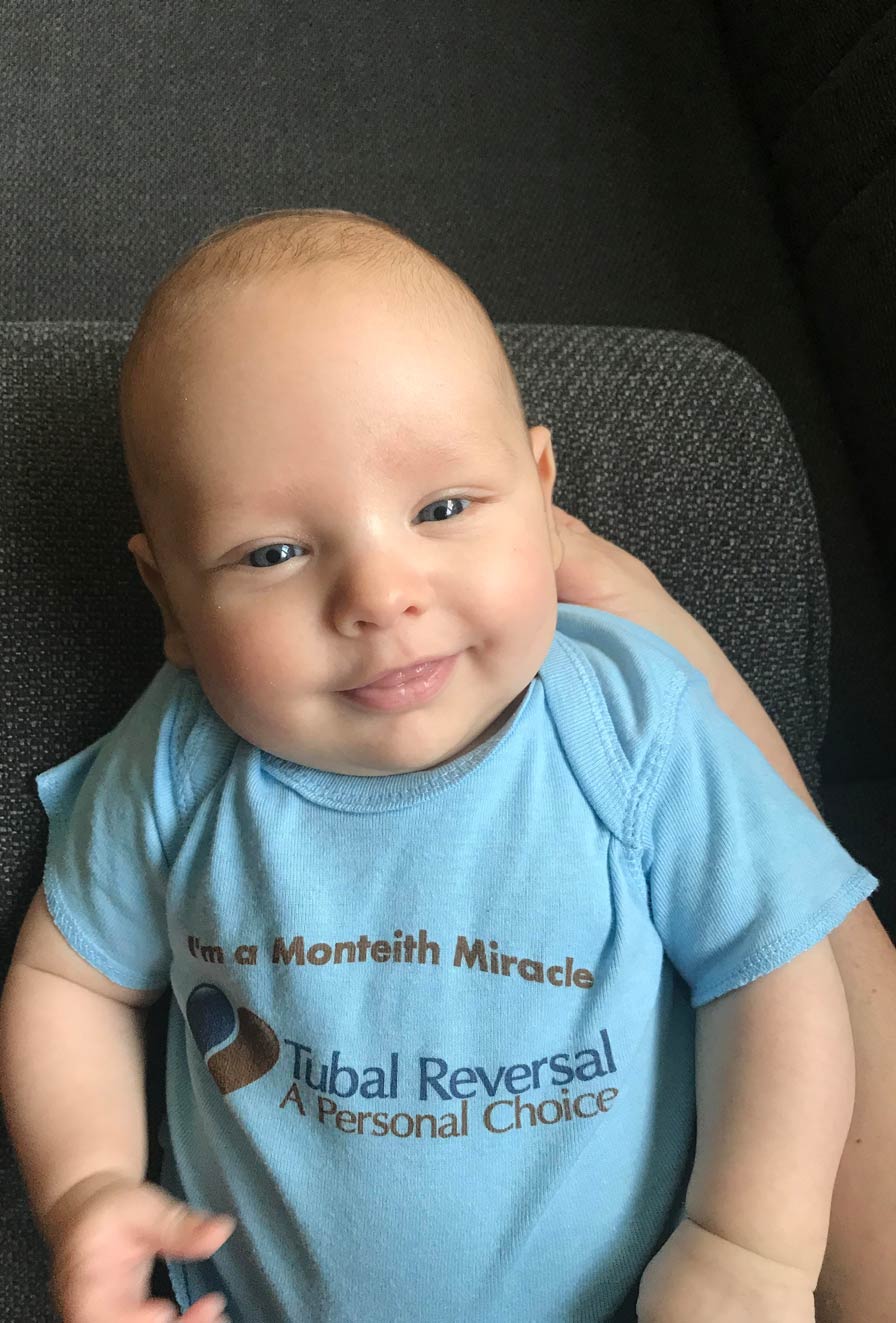
“Ivy is our second Monteith Miracle!”
– APO, Armed Forces Europe
 Birth Announcements
“It was a leap of faith and we have had a blessed journey.”
– Colorado Springs, Colorado
 Birth Announcements
“Thank you for making our dreams come true!”
– Nebo, North Carolina
We want you to update us on your progress after surgery at our center. Your feedback is important to us!
Pregnancy Report Form
Update Us About Your Pregnancy
Birth Report Form
Your questions about reversal surgery will be answered directly and promptly by Dr. Monteith. Please read our most frequently asked questions before submitting your questions. Dr. Monteith will not respond to general medical questions or if you are a patient of another doctor and are contacting him because your doctor is unavailable.* If you have been a patient at our reversal center and have specific questions about your medical care you should contact the reversal staff directly at (919) 977-5050.

