Removing Essure Devices
Manual traction is feasible
Removing Essure devices intact can be done with manual traction. In our experience, we have found this is most easily done within the first several months of insertion and before the inserts become locked in place from fibroelastic tissue.
With gradual in-growth of fibroelastic tissue, removing Essure micro-inserts will become increasingly more difficult to remove when using manual traction alone.
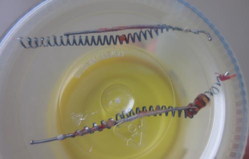
The lower micro-insert was in the uterine cavity.
The upper micro-insert was in the isthmic section of the fallopian tube. The lateral end of the inner coil near the spherical radiographic marker shows distortion in the area where it was manipulated with a surgical hemostat.
Removing Essure Devices: Traction works soon after insertion
The Polyethylene terephthalate (PET) fibers are wrapped around the stainless steel inner coil. These fibers cause reactive fibrosis. This is a gradually process which results in scar tissue in-growth into the Essure micro-inserts.
As fibrosis occurs the Essure devices become more secure in their position. As the scar tissue in-growth occurs, the devices become progressively locked into position and more force is required for extraction.
This photo demonstrates Essure devices we were able to remove successfully and intact using manual traction.
This patient underwent Essure sterilization approximately 8 weeks prior to requesting removal. She experienced generalize pain, dysmenorrhea, and vaginal bleeding. At the time of her removal one coil was found to be completely in the cavity of her uterus and the second coil was in the proper location. The intrauterine micro-insert was removed with a vaginal approach using a polyp forceps. The intratubal micro-insert was removed easily through an incision in the tube and manual traction.
Tissue in-growth makes Removing Essure devices more difficult
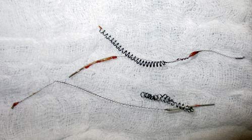
Micro-inserts which required significant traction for removal. Note the wire like appearance of the stretched inner coil. The outer coil will often retain its coil-like shape regardless of the amount of traction required for removal.
The appearance of the micro-inserts provides a visual clue as to how much fibrous tissue in-growth was present and how much traction was required when removing Essure devices.
The longer the coils have remained in the tubes then the more in-growth of scar tissue occurs and the more traction that needs to be applied during removal.
The inner coil is stainless steel and is tightly wound around the PET fibers. The more traction required on this coil results in the inner coil becoming unwound and assuming a more wire like in appearance. The inner coil seems significantly stronger and less prone to fracture than the outer coil.
The outer coil is nitinol and this coil will often resume its shape after removal regardless of the amount of traction. The outer coil seems to have less tensile strength and more prone to fracture when excessive traction has been applied.
When the inner coil retains its coil-like state then less force was required for removing Essure devices. When the inner coil is unwound and wire-like then more traction was required to remove Essure devices.
